
Introduction
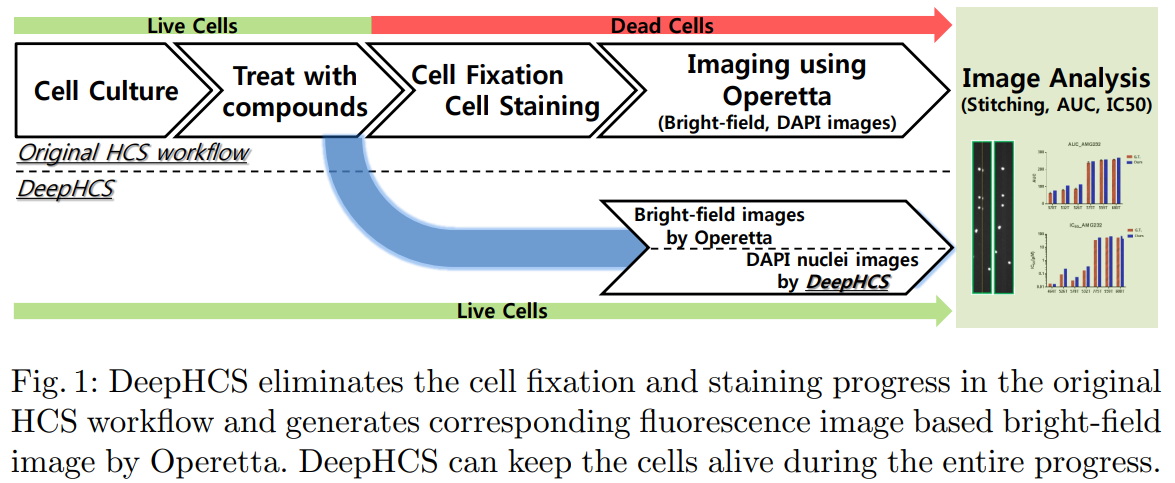
A glioblastoma (GBM) is a brain tumor that is commonly found in the cerebral hemisphere of the brain. GBM is considered an obstinate brain tumor because even after medical advances in the past few decades, no effective treatment has been discovered that greatly improves life expectancy in patients. The patient-specific chemotherapy by analyzing patient-driven GBM tumor cells to find the most effective drug for the target patient, called precision medicine, has become popular. High-throughput screening (HCS) uses high-throughput imaging and automatic image analysis to evaluate changes in the phenotype of the whole cells, such as counting the number of living cells versus dead cells, measuring the size of the cells, comparing the shape of the cells, etc.
Motivation
There have been many research efforts to develop image processing techniques for bright-field imaging to extract cell phenotypes without fluorescence imaging. However, most previous works focused only on cell segmentation and detection directly from bright-field images, and no state-of-the-art deep learning methods are leveraged. In addition, in spite of ongoing research efforts in bright-field image analysis, the standard HCS workflow still relies on detecting and analyzing biomarkers presented in fluorescence images.
Based on these observations, we propose DeepHCS, a novel data-driven image conversion technique for high-content screening.
DeepHCS : Bright-field to Fluorescence Microscopy Image Conversion using Deep Learning for Label-free High-Content Screening
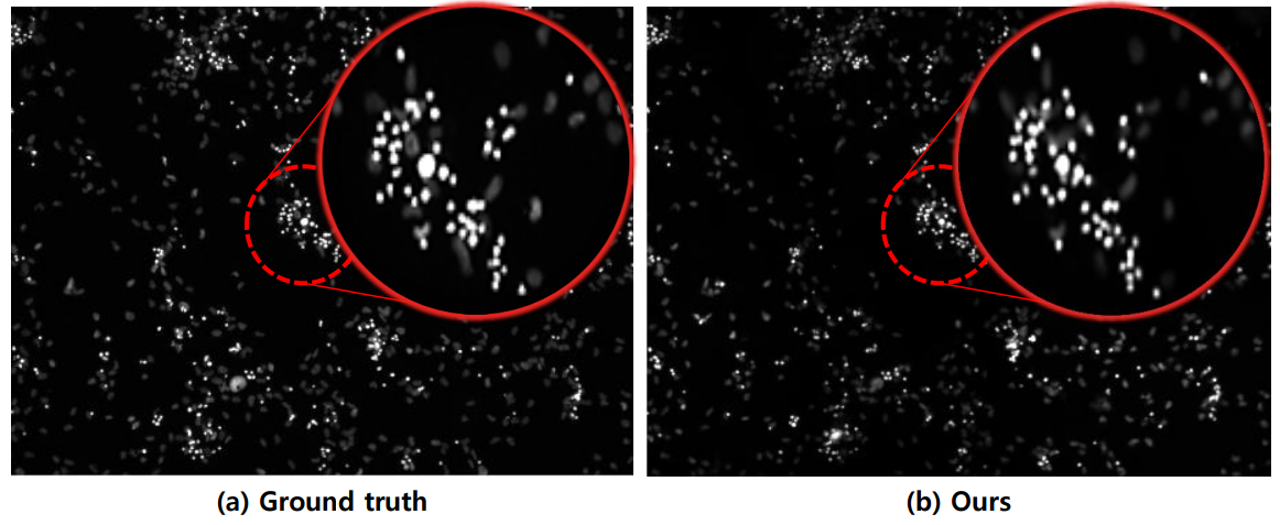
The main motivation of the proposed work is to automatically generate virtual biomarker images from conventional bright-field images, which can greatly reduce time-consuming and laborious tissue preparation efforts and improve the throughput of the screening process. By leveraging a state-of-the-art deep learning method, the proposed method can produce synthetic fluorescence images comparable to real DAPI images with high accuracy.
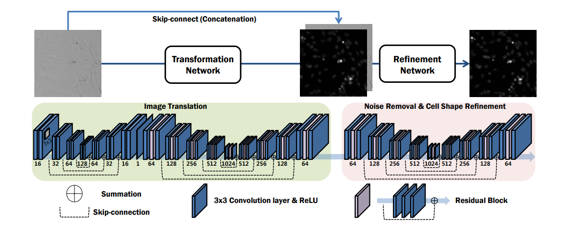
DeepHCS uses bright-field images and their corresponding cell nuclei staining (DAPI) fluorescence images as a set of image pairs to train a series of end-to-end deep convolutional neural networks.
Results

[bibtex file=ghlee_deephcs_2018.bib format=plain
process_titles=0]